


Insana Lab: Ultrasonic Imaging - The University of Illinois at Urbana-Champaign
Insana Lab: Ultrasound Research Interface (URI)
Tutorials | B-Mode | 2D Color Doppler | More Color Doppler | Scan Conversion | M-mode | Spectral Doppler | Misc. FunctionsScan Conversion
Scan conversion allows for accurate display of sector or parallelogram images from RF data.
B and C image data are stored and displayed on a rectangular grid unless the scan converter algorithm is applied.
The B-mode scan-conversion is done automatically when "1" is entered in the "Display scan-conversion?" field of the prompt window of URIBmode.m;
the color flow displays are formed by pressing the "Full FOV" button on the GUI of URICBmode.m.
Three basic types of array transducers are used to acquire different sector and parallelogram images:
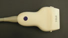 Linear Array (and linear-phased array): rectangular, steered parallelogram, and sector images. Linear Array (and linear-phased array): rectangular, steered parallelogram, and sector images.
|
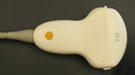 Curved Array (also known as convex or curvilinear array): sector images. Curved Array (also known as convex or curvilinear array): sector images.
|
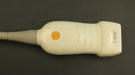 Phased Array (also known as sector array): sector images. Phased Array (also known as sector array): sector images.
|
The following images are examples of scan-conversion in different imaging modes and transducers.
|
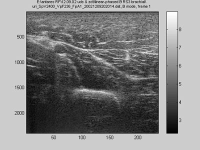
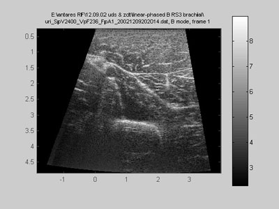
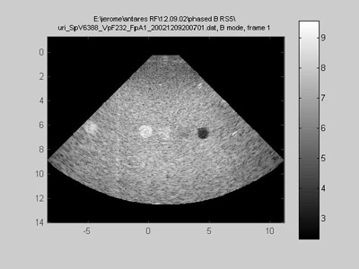
Linear-phased array (B-mode) cross-section of upper arm (including brachial artery, biceps, brachialis, triceps, and humerus bone) |
||
|---|---|---|
 |

|
 |
|
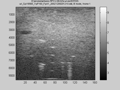
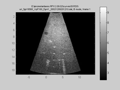
Curved (convex) array (B-mode) calibration phantom |
||
 |
|
 |
|
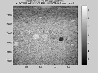
Phased array (B-mode) calibration phantom |
||
 |
|
 |
|
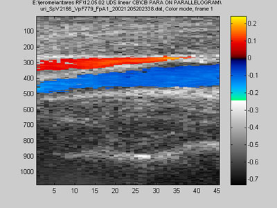
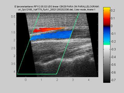
Steering with linear array (2D color flow) longitudinal view of carotid artery |
||
 |
|
 |
|
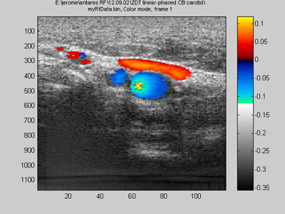
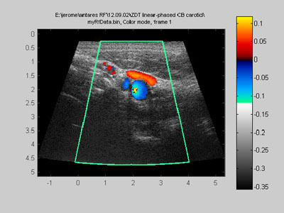
Linear-phased array (2D color flow) cross-sectional view of carotid artery |
||
 |
|
 |
