


Insana Lab: Ultrasonic Imaging - The University of Illinois at Urbana-Champaign
Project 1: Elasticity Imaging of Breast Cancer
5.5.05
|
Fig 1. Elasticity Imaging is a diagnostic technique for noninvasive visualization of soft tissue stiffness. Elasticity imaging can detect disease processes involving local inflammatory responses, which can be acute or chronic and associated with fibrosis, hyperplasia, and neoplasia. Elasticity methods is also palpation (Fig 1), however in place of the fingers standard imaging methods are used to remotely sense the deformation patterns resulting from an applied force. The advantage over manual palpation is high depth resolution and increase sensitivity to deep lesions. The term "elasticity imaging" encompasses a large range of techniques, although many are based on imaging deformation or strain. |
|
|
|
|
|
|
|
|
|
|
 0.5) and elastic. Small applied stresses ensure that the stress-strain curve is linear.
However if we hold the stress constant longer than a few seconds, we see that the strain
increases slowly over time (see plots in Fig 5). Whether we are imaging water-based gelatin
or breast tissue, it seems there are two physical mechanisms behind the mechanical relaxation.
These are related to fluid flow (poroelasticity) and cross-link relaxation (viscoelasticity).
Therefore strains measured many seconds after applying and holding a stress (during the solid
phase of the deformation process in Fig 5) will react as compressible viscoelastic solids.
Since we are applying a "known" stress and measuring the local strain, the bi-exponential curves
of strain increase over time measured at each pixel location (see Figs 5 – 7) is given by the
classical Kelvin-Voigt model (see Fig 7b and [6]). 0.5) and elastic. Small applied stresses ensure that the stress-strain curve is linear.
However if we hold the stress constant longer than a few seconds, we see that the strain
increases slowly over time (see plots in Fig 5). Whether we are imaging water-based gelatin
or breast tissue, it seems there are two physical mechanisms behind the mechanical relaxation.
These are related to fluid flow (poroelasticity) and cross-link relaxation (viscoelasticity).
Therefore strains measured many seconds after applying and holding a stress (during the solid
phase of the deformation process in Fig 5) will react as compressible viscoelastic solids.
Since we are applying a "known" stress and measuring the local strain, the bi-exponential curves
of strain increase over time measured at each pixel location (see Figs 5 – 7) is given by the
classical Kelvin-Voigt model (see Fig 7b and [6]).
|
|
|
|
|
|
Fig 7a shows there are three terms result from a step stress stimulus. The first is These are illustrated in Fig 7a where, in this example, the inclusion is different from the background because of a higher collagen concentration. |
|
|
|
|
|
|
|
|
Fig 9. We have been conducting human subject trials at UD Davis Medical Center with advice and guidance from Dr Karen Lindors MD, chief of the Breast Clinic. Examples are shown in Fig 9. Our patient population is different from other studies in that we select nonpalpable lesions from patients with suspicious mammograms that are undergoing US guided biopsy. We have pathological reports on each case to establish a diagnosis. Fig 9 shows three nonpalpable lesions that appear, left-to-right, stiffer (darker in strain) than the background, the same stiffness as the background, and softer (brighter in strain) than the background. Whereas palpable lesions are most often stiff, NP lesions can present with a variety of features. Does this finding lessen the value of elasticity imaging for breast lesion classification? I think not, but it does mean that we need more information to improve specificity. |
|
|
|
|
|
|
|
|
|
|
|
|
|
|
|
|
 to increase. This is precisely what we see in
Fig 17, showing breast images from a patient
with a benign lesion. The variation in strain with time due to interstitial fluid movement is known as
poroelasticity. The mechanics of porous media was developed by Biot in a series of papers [15] beginning
in 1941 to describe soil consolidation under a load. These concepts have been expanded recently to modeling
[16] particularly of cartilage dynamics [17] and other biological tissues. to increase. This is precisely what we see in
Fig 17, showing breast images from a patient
with a benign lesion. The variation in strain with time due to interstitial fluid movement is known as
poroelasticity. The mechanics of porous media was developed by Biot in a series of papers [15] beginning
in 1941 to describe soil consolidation under a load. These concepts have been expanded recently to modeling
[16] particularly of cartilage dynamics [17] and other biological tissues.
|
|
|
for malignant lesions as in Fig 18. Consequently, it appears to be helpful
to measure time-varying strain and the associated relaxation constant
to clearly differentiate
malignant from benign lesions.
|
|
|
|
|
|
|
|
 ,
is due to the relaxation of
hydrogen- and electrostaticaly-bonded cross-links within collagen fibers, Fig 19. Covalent
bonding between microfibrils is sparse in youth but increases slowly in organisms with age.
They are robust to mechanical and thermal stress and most other stimuli encountered in vivo.
The covalent bonds are most likely to be the source of the solid phase strain. That is, if we
let ,
is due to the relaxation of
hydrogen- and electrostaticaly-bonded cross-links within collagen fibers, Fig 19. Covalent
bonding between microfibrils is sparse in youth but increases slowly in organisms with age.
They are robust to mechanical and thermal stress and most other stimuli encountered in vivo.
The covalent bonds are most likely to be the source of the solid phase strain. That is, if we
let  in (1), the strain becomes in (1), the strain becomes 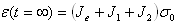 .
Strain is proportional to the sum of reactances [6] from each
component of the mechanical response. Note that the shear modulus in the solid phase is .
Strain is proportional to the sum of reactances [6] from each
component of the mechanical response. Note that the shear modulus in the solid phase is 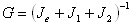 . However,
between the time that the step stress is applied and the strain reaches equilibrium, . However,
between the time that the step stress is applied and the strain reaches equilibrium,  , the fragile
hydrogen-bonded cross-links are released to lower internal stresses. They will then reform but at
a lower energy state. We have begun examining this mechanism as a method for imaging
metabolically-driven changes in extracellular pH [19]. More on this later. , the fragile
hydrogen-bonded cross-links are released to lower internal stresses. They will then reform but at
a lower energy state. We have begun examining this mechanism as a method for imaging
metabolically-driven changes in extracellular pH [19]. More on this later.
|
|
|
1.C Gelatin as a Physical Model for Imaging Development
Pure, natural gelatin is collagen. Formation begins as polypeptide fragments of a few types of amino acids (three most common are glycine, proline, and hydroxyprolin, Fig 18). The fragments are joined through peptide linkages to assemble into a triple helix structure [20]. When added to water at the correct temperature and concentration, it forms a gel. Commercially available gelatin powder is produced from denatured collagen derived, in most common uses, from fibrous animal tissues like skin [21]. Impure forms of gelatin, including commercial lab-grades, are distinguishable as gelatine. Like breast stroma, gelatin contains mostly type I collagen molecules, but few fibers are formed in network. The network is a hydrocolloid (water-loving) material that absorbs up to 10 times its mass in water. Unlike stromal tissue, in vivo, there is no polysaccharide gel surrounding the collagen fibers. The few fibers that form are shorter in length depending on pH, temperature history and other environmental factors, and the cross-linking is less robust than that in vivo. Nevertheless, gelatinous collagen fibers expose electrically charged sites that adsorb and structure water in a manner similar to the polysaccharide gels in tissue. Also aldehydes and other chemical cross-linking agents are used to temporally and thermally stabilize the colloid structure. Like connective tissues, in vivo, the number of covalent bonds between collagen fibers in gelatin increase as the gel ages. There is literature on the shear and bulk elastic properties of gelatin used to develop ultrasound phantoms for medical imaging applications from group at University of Wisconsin and elsewhere [22-24]. However, the study of poro- and viscoelastic responses is predominantly in the vast literature on food sciences and medical prosthetic devices. Gelatin is usually produced from a process that denatures collagenous biomaterials by treatment with alkali or acidic solutions [20,21]. Type A gelatin uses acidic processing, and type B gelatin uses alkali processing. Gelatin type is extremely important if you are interested in time-varying mechanical responses, because type determines the isoelectric point. The isoelectric point (IEP) of a gel is the pH of a solution containing gelatin proteins whereby the following measurement properties are all minimized: movement of particles in an electric field, conductivity, solubility, osmotic pressure, swelling, and viscosity [25]. In short, IEP is the pH where the gel is least reactive (most stable) with respect to the measurements listed above. This is not to be confused with the isoionic point (IIP) [25], which is defined as the pH of a solution for which the addition of gelatin causes no change in the pH value of the solution.1 If the gelatin is pure collagen protein and not gelatine, as are most lab-grade products, then IEP = IIP. In that special case, IIP is the pH of a solution where there is no net charge on the surface of the collagen fibers [21]. IIP for acid-precursor gelatins (type A) is in a pH range of 9.0-9.2, similar to native collagen [20]. IIP for alkali-precursor gelatins (type B) is in a pH range of 4.8 – 5.2. However IEP can be most anywhere on the pH scale; the density of charged sites depends in how the gelatin was prepared and its purity. Gelatin viscosity will double as the net molecular charge per collagen molecule increases from 0 at IEP to ±30 (see Fig II-30 in [20]). 1 There seem to be an endless supply of definitions for IEP and IIP, and, not being a biochemist, I am unsure how to interpret most of them. For pure clarity of prose, I prefer the discussion by Adair from 1937 [25]. |
|
|
References:
[1] Chaturvedi P, Insana MF, Hall TJ: 2-D companding for noise reduction
in strain imaging. IEEE Trans. Ultrason. Ferro. Freq. Contrl.
UFFC-45:179-191, 1998. |
 and is constant with
time; it describes strain during the incompressible phase, before relaxation can occur. When measurement
time
and is constant with
time; it describes strain during the incompressible phase, before relaxation can occur. When measurement
time  is very close to the time that the stress was applied
  , then
 . This is the quantity we normally
think of as a strain image in static elasticity imaging. The second two terms in (1) grow in amplitude
over time. The amount of growth depends on the elastic constants
. This is the quantity we normally
think of as a strain image in static elasticity imaging. The second two terms in (1) grow in amplitude
over time. The amount of growth depends on the elastic constants  and
and
 , and the corresponding rates of
growth are characterized by time constants
, and the corresponding rates of
growth are characterized by time constants  ,
,